Catatonia
Session: Poster Session
(017) Evaluating and Reducing Risk of Electroconvulsive Therapy in the Presence of Obstructive Hydrocephalus: A Case Report

Trainee Involvement: Yes

Karanbir Padda, MD
Assistant Professor of Clinical Psychiatry
New York-Presbyterian/Weill Cornell Medical Center
Brooklyn, New York, United States- KQ
Kelvin Quinones-Laracuente, MD, PhD
Psychiatry resident
NYU
New York, New York, United States - JL
Justin Lewin, MD
Consultation-Liaison Psychiatry Attending
Bellevue Hospital
New York, New York, United States
Presenting Author(s)
Co-Author(s)
References: Weiner R et al. The practice of electroconvulsive therapy, second edition. American Psychiatric Association, 2001. Hermida AP, Job G, Tang Y. Electroconvulsive therapy in a geriatric patient with normal pressure hydrocephalus without a shunt. J ECT. 2014. Patkar AA, Hill KP, Weinstein SP, Schwartz SL. ECT in the presence of brain tumor and increased intracranial pressure: evaluation and reduction of risk. J ECT. 2000 Jun;16(2):189-97.
Background: Per the American Psychiatric Association, there are no absolute contraindications to electroconvulsive therapy (ECT). Elevated intracranial pressure (ICP) is considered a relative contraindication (Weiner et al., 2001). We present a case of a patient suffering from catatonia with comorbid obstructive hydrocephalus. We explore the risks of ECT in the context of obstructive hydrocephalus and propose a novel neurosurgical strategy to reduce the risk of elevated ICP.
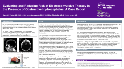
Case: A 65-year-old male with intellectual disability, major depressive disorder, panhypopituitarism, and severe congenital hydrocephalus secondary to aqueductal stenosis was admitted to a tertiary center for catatonia (Bush Francis Catatonia Rating Scale = 18). The patient was treated with IV lorazepam, but there was minimal improvement. The option of ECT was explored with multiple consulting teams, including psychiatry, medicine, neurology, neurosurgery, and ophthalmology. The patient’s MRI brain and ophthalmologic exam indicated chronically elevated ICP. Due to the risks of brain herniation and iatrogenic subdural hemorrhage, lumbar puncture and ventriculoperitoneal shunt as means to relieve ICP were deemed too dangerous. A plan was proposed to place an external ventricular drain (EVD) to monitor ICP during ECT. If ICP does not increase during the first ECT session, the EVD will be removed, and ECT can be continued safely without further monitoring. If ICP increases during ECT, the EVD will remain in place throughout the course of ECT to drain cerebrospinal fluid and relieve ICP to mitigate risk of neurologic complications.
Discussion: Our case report is the first to explore the use of ECT in a patient with obstructive hydrocephalus. ECT has successfully and safely been performed in patients with normal pressure hydrocephalus (Hermida et al., 2014). However, obstructive hydrocephalus presents the issue of elevated ICP. ECT is thought to elevate ICP, so strategies to relieve ICP are necessary to prevent neurologic complications. In a previous case, ECT was performed in the setting of a brain tumor and elevated ICP, utilizing dexamethasone to decrease brain edema and reduce ICP (Patkar et al., 2000). We propose a strategy of using an EVD to monitor and relieve ICP to mitigate risk of performing ECT in the setting of obstructive hydrocephalus. Further research is needed into the safety of ECT and risk-mitigating strategies in the setting of obstructive hydrocephalus.
Conclusion: With risk-mitigating strategies, ECT may be considered a treatment option in the presence of obstructive hydrocephalus and elevated ICP.

